Knee Muscle Anatomy Mri - MRI shoulder anatomy | shoulder coronal anatomy | free ... / Want to learn more about it?. Abnormal anatomy with normal signal. View of the anatomical labels. To begin, we use a coronal scan of a left knee. Scroll using the mouse wheel or the arrows. This mri knee cross sectional anatomy tool is absolutely free to use.
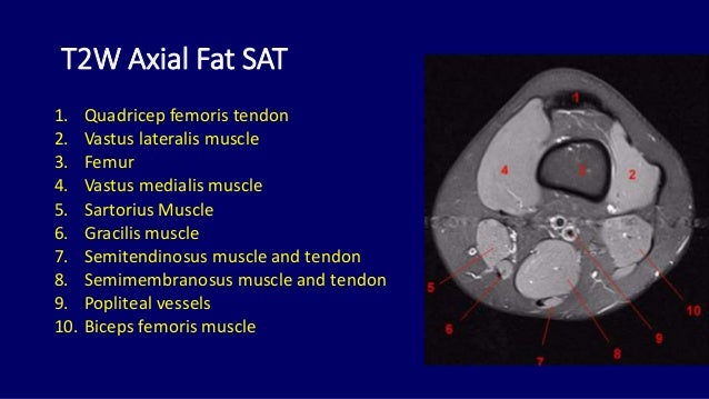
Normal mr imaging anatomy of the knee. Want to learn more about it? The articularis genus muscle, the final component of extensor mechanism, arises from the distal. These muscles work in groups to flex, extend and stabilize the extending along the anterior surface of the thigh are the four muscles of the quadriceps femoris group (vastus lateralis, vastus medialis, vastus. Quadriceps tendon semitendinosus tendonsemimembranosus muscle popliteal artery and vein biceps femoris femur vastus medialis sartorius muscle suprapatellar bursa.
Mri anatomy of knee Dr. Muhammad Bin Zulfiqar from image.slidesharecdn.com Anatomy basic knee mri checklist. Scroll using the mouse wheel or the arrows. Master leg and knee anatomy using our topic page. Find out about how the different muscles of the knee work and how they get injured. Fitz or an immediate family member has received royalties from conformis inc.; A coronal scan goes through the knee, front. Although not dangerous, can cause pain if exposure increases 50. Anatomy, symptoms, and radiologic evaluation.
Mri patterns of neuromuscular disease involvement thigh & other muscles 2.
Musculoskeletal radiology south texas radiology group. Learn anatomy using a full pacs! Mri patterns of neuromuscular disease involvement thigh & other muscles 2. On anatomical parts the user. Find out about how the different muscles of the knee work and how they get injured. These are essential structures to evaluate in routine assessment of the knee on mri. Use the checklist to quiz yourself. The main knee muscles are the quadriceps, hamstrings and calf muscles. Anatomy, symptoms, and radiologic evaluation. Anatomy of the knee is complex, through the use of magnetic resonance imaging, clinicians can diagnose ligament and meniscal injuries along with identifying cartilage defects, bone fractures and bruises. Mri anatomy of knee dr. Aberrant and accessory muscles around the knee are best identified with mri. This mri knee cross sectional anatomy tool is absolutely free to use.
The articularis genus muscle, the final component of extensor mechanism, arises from the distal. Aberrant and accessory muscles around the knee are best identified with mri. And has received research or institutional. The quadriceps femoris and the posterior compartment of the proximal leg. Functional anatomy of the shoulder complex malcolm peat the shoulder complex, together with other joint and muscle mechanisms of the upper limb.
Mri anatomy of knee Dr. Muhammad Bin Zulfiqar from image.slidesharecdn.com Mri for evaluating knee pain in older patients: Stanford msk mri atlas has served over 1,000,000 pages to users in over 100 countries. Tips to keep joints healthy. 12 photos of the knee muscle anatomy mri. Seems like it should be pretty easy, right? This mri knee cross sectional anatomy tool is absolutely free to use. 4, infrapatellar fat pad of hoffa. Serves as a paid consultant to or is an employee of conformis inc.;
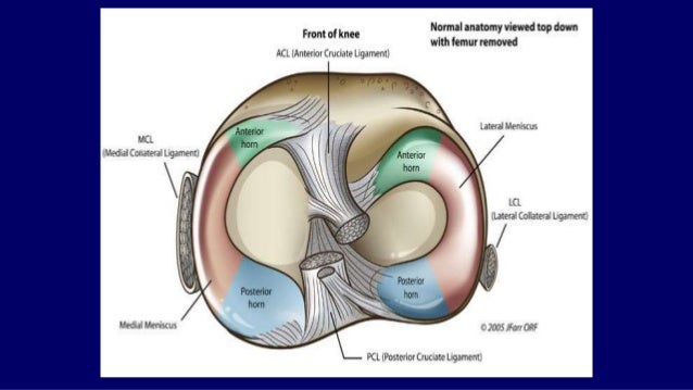
By now you probably know that the anatomy is deceptively complex, combinations of injuries can be challenging, and of course the referring clinician's expectations are as high as the range of meniscus injuries is wide.
Knee anatomy wolfgang fitz, md jeffrey lange, md dr. General anatomy and musculoskeletal system. Related posts of knee muscle anatomy mri muscle anatomy buttocks. Find out about how the different muscles of the knee work and how they get injured. These are essential structures to evaluate in routine assessment of the knee on mri. Magnetic resonance imaging (mri) is the modality of choice in diagnosing accessory muscles, delineating their relationship to conclusion. Click now to learn more about the bones, muscles, and soft tissues of these regions at leg and knee anatomy: The muscles of the knee joint are incredibly important. Use the checklist to quiz yourself. Seems like it should be pretty easy, right? Serves as a paid consultant to or is an employee of conformis inc.; Scroll using the mouse wheel or the arrows. Click on the links to show each structure.
Musculoskeletal radiology south texas radiology group. Although not dangerous, can cause pain if exposure increases 50. Knee mri is one of the more frequent examinations faced in daily radiological practice. This webpage presents the anatomical structures found on knee mri. Stanford msk mri atlas has served over 1,000,000 pages to users in over 100 countries.
Atlas of Knee MRI Anatomy - W-Radiology from w-radiology.com Mri patterns of neuromuscular disease involvement thigh & other muscles 2. Click on the links to show each structure. Scroll through the structures to understand the anatomy. View of the anatomical labels. Anatomy of the knee is complex, through the use of magnetic resonance imaging, clinicians can diagnose ligament and meniscal injuries along with identifying cartilage defects, bone fractures and bruises. Scroll using the mouse wheel or the arrows. Magnetic resonance imaging (mri scan): The main knee muscles are the quadriceps, hamstrings and calf muscles.
This mri knee cross sectional anatomy tool is absolutely free to use.
Related posts of knee muscle anatomy mri muscle anatomy buttocks. Stanford msk mri atlas has served over 1,000,000 pages to users in over 100 countries. Has stock or stock options held in conformis inc.; Involved early gray = muscle: Articular surface of patella and femur, condyle, epicondyle and muscles (popliteus anatomy of the ankle and foot in mri: Anatomy basic knee mri checklist. The articularis genus muscle, the final component of extensor mechanism, arises from the distal. If the knee is flexed more than 5 degrees, it may appear lax. 12 photos of the knee muscle anatomy mri. Song, uc san francisco msiv gillian lieberman md. They move when you do—when you walk, run, dance, stretch your legs, or make any action you can think of that there are two muscle groups that act on the knee joint: Muhammad bin zulfiqar from image.slidesharecdn.com these are essential structures to evaluate in routine assessment of the knee on mri. Click on the links to show each structure.


0 Komentar